
 Datasheet
 MSDS
MSDSDescription:Rabbit polyclonal antibody to Oncostatin MImmunogen:Recombinant fusion protein of human Oncostatin M. The exact sequence is proprietary.Purification:The antibody was purified by immunogen affinity chromatography.Clonality:PolyclonalForm:Liquid in 0.42% Potassium phosphate, 0.87% Sodium chloride, pH 7.3, 30% glycerol, and 0.01% sodium azide.Dilution:WB (1/500 - 1/1000), IF/IC (1/50 - 1/200)Gene Symbol:OSMAlternative Names:Oncostatin-M; OSM
Entrez Gene (Human):
5008;
Entrez Gene (Mouse):
18413;
Entrez Gene (Rat):
289747;
SwissProt (Human):
P13725;
SwissProt (Mouse):
P53347;
SwissProt (Rat):
Q65Z15;
Storage/Stability:Shipped at 4°C. Upon delivery aliquot and store at -20°C for one year. Avoid freeze/thaw cycles.
-
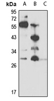 Western blot analysis of Oncostatin M expression in HepG2 (A), NIH3T3 (B), mouse heart (C) whole cell lysates. (Predicted band size: 28 kD; Observed band size: 28 kD)
 -
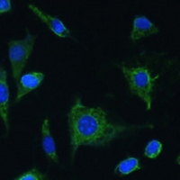 Immunofluorescent analysis of Oncostatin M staining in L929 cells. Formalin-fixed cells were permeabilized with 0.1% Triton X-100 in TBS for 5-10 minutes and blocked with 3% BSA-PBS for 30 minutes at room temperature. Cells were probed with the primary antibody in 3% BSA-PBS and incubated overnight at 4 °C in a hidified chamber. Cells were washed with PBST and incubated with a Alexa Fluor 488-conjugated secondary antibody (green) in PBS at room temperature in the dark.