
 Datasheet
 MSDS
MSDSDescription:Rabbit polyclonal antibody to MFN1Immunogen:Recombinant full length protein of human MFN1Purification:The antibody was purified by immunogen affinity chromatography.Clonality:PolyclonalForm:Liquid in 0.42% Potassium phosphate, 0.87% Sodium chloride, pH 7.3, 30% glycerol, and 0.01% sodium azide.Dilution:WB (1/500 - 1/2000), IF/IC (1/50 - 1/200)Gene Symbol:MFN1Alternative Names:Mitofusin-1; Fzo homolog; Transmembrane GTPase MFN1
Entrez Gene (Human):
55669;
Entrez Gene (Mouse):
67414;
Entrez Gene (Rat):
192647;
SwissProt (Human):
Q8IWA4;
SwissProt (Mouse):
Q811U4;
SwissProt (Rat):
Q8R4Z9;
Storage/Stability:Shipped at 4°C. Upon delivery aliquot and store at -20°C for one year. Avoid freeze/thaw cycles.
-
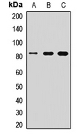 Western blot analysis of MFN1 expression in A549 (A), mouse liver (B), rat brain (C) whole cell lysates. (Predicted band size: 41; 71; 84 kD; Observed band size: 84 kD)
 -
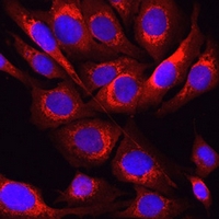 Immunofluorescent analysis of MFN1 staining in U2OS cells. Formalin-fixed cells were permeabilized with 0.1% Triton X-100 in TBS for 5-10 minutes and blocked with 3% BSA-PBS for 30 minutes at room temperature. Cells were probed with the primary antibody in 3% BSA-PBS and incubated overnight at 4 °C in a humidified chamber. Cells were washed with PBST and incubated with a AF594-conjugated secondary antibody (red) in PBS at room temperature in the dark. DAPI was used to stain the cell nuclei (blue).
Differential Expression of Lonp1 Isoforms in Cancer Cells