
 Datasheet
 MSDS
MSDSDescription:Mouse monoclonal antibody to Peroxiredoxin 1Immunogen:Recombinant protein corresponding to human Peroxiredoxin 1.Purification:Affinity chromatographyClonality:MonoclonalForm:Liquid in 0.42% Potassium phosphate, 0.87% Sodium chloride, pH 7.3, 30% glycerol, and 0.01% sodium azide.Dilution:WB (1/1000 - 1/3000), IF/IC (1/100 - 1/200)Gene Symbol:PRDX1Alternative Names:PAGA; PAGB; TDPX2; Peroxiredoxin-1; Natural killer cell-enhancing factor A; NKEF-A; Proliferation-associated gene protein; PAG; Thioredoxin peroxidase 2; Thioredoxin-dependent peroxide reductase 2
Entrez Gene (Human):
5052;
Entrez Gene (Mouse):
18477;
Entrez Gene (Rat):
117254;
SwissProt (Human):
Q06830;
SwissProt (Mouse):
P35700;
SwissProt (Rat):
Q63716;
Storage/Stability:Shipped at 4°C. Upon delivery aliquot and store at -20°C for one year. Avoid freeze/thaw cycles.
-
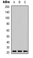 Western blot analysis of Peroxiredoxin 1 expression in MCF7 (A), mouse brain (B), rat kidney (C) whole cell lysates. (Predicted band size: 22 kD; Observed band size: 21 kD)
 -
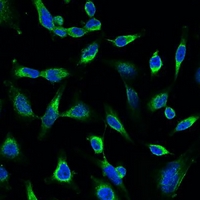 Immunofluorescent analysis of Peroxiredoxin 1 staining in Hela cells. Formalin-fixed cells were permeabilized with 0.1% Triton X-100 in TBS for 5-10 minutes and blocked with 3% BSA-PBS for 30 minutes at room temperature. Cells were probed with the primary antibody in 3% BSA-PBS and incubated overnight at 4 °C in a hidified chamber. Cells were washed with PBST and incubated with a FITC-conjugated secondary antibody (green) in PBS at room temperature in the dark. DAPI was used to stain the cell nuclei (blue).