
 Datasheet
 MSDS
MSDSDescription:Rabbit polyclonal antibody to IGF1 Receptor (Phospho-Y1161)Immunogen:KLH-conjugated synthetic phosphopeptide corresponding to residues surrounding Y1161 of human IGF1 Receptor protein. The exact sequence is proprietary.Purification:The antibody was purified by immunogen affinity chromatography.Clonality:PolyclonalForm:Liquid in 0.42% Potassium phosphate, 0.87% Sodium chloride, pH 7.3, 30% glycerol, and 0.01% sodium azide.Dilution:WB (1/500 - 1/1000), IH (1/100 - 1/200), IF/IC (1/100 - 1/500), IP (1/10 - 1/100)Gene Symbol:IGF1RAlternative Names:IGF1R; Insulin-like growth factor 1 receptor; Insulin-like growth factor I receptor; IGF-I receptor; CD221; INSR; Insulin receptor; IR; CD220
Entrez Gene (Human):
3480;
3643;
Entrez Gene (Mouse):
16001;
16337;
Entrez Gene (Rat):
25718;
SwissProt (Human):
P08069;
P06213;
SwissProt (Mouse):
Q60751;
P15208;
SwissProt (Rat):
P24062;
P15127;
Storage/Stability:Shipped at 4°C. Upon delivery aliquot and store at -20°C for one year. Avoid freeze/thaw cycles.
-
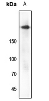 Western blot analysis of IGF1 Receptor (Phospho-Y1161) expression in zebrafish (A) whole cell lysates. (Predicted band size: 154; 156 kD; Observed band size: 200 kD)
 -
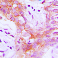 Immunohistochemical analysis of IGF1 Receptor (Phospho-Y1161) staining in human breast cancer formalin fixed paraffin embedded tissue section. The section was pre-treated using heat mediated antigen retrieval with sodium citrate buffer (pH 6.0). The section was then incubated with the antibody at room temperature and detected using an HRP conjugated compact polymer system. DAB was used as the chromogen. The section was then counterstained with haematoxylin and mounted with DPX.
 -
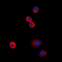 Immunofluorescent analysis of IGF1 Receptor (Phospho-Y1161) staining in HeLa cells. Formalin-fixed cells were permeabilized with 0.1% Triton X-100 in TBS for 5-10 minutes and blocked with 3% BSA-PBS for 30 minutes at room temperature. Cells were probed with the primary antibody in 3% BSA-PBS and incubated overnight at 4 °C in a humidified chamber. Cells were washed with PBST and incubated with a DyLight 594-conjugated secondary antibody (red) in PBS at room temperature in the dark. DAPI was used to stain the cell nuclei (blue).
The insulin-like growth factor pathway is altered in spinocerebellar ataxia type 1 and type 7
AF4 is a critical regulator of the IGF-1 signaling pathway during Purkinje cell development
High IGF-IR activity in triple-negative breast cancer cell lines and tumorgrafts correlates with sensitivity to anti-IGF-IR therapy
Upregulation of insulin-like growth factor binding protein 3 in astrocytes of transgenic mice that express Borna disease virus phosphoprotein
Igf1r signaling is indispensable for preimplantation development and is activated via a novel function of E-cadherin
Lkb1/Stk11 regulation of mTOR signaling controls the transition of chondrocyte fates and suppresses skeletal tumor formation
Insulin-like growth factor 1 receptor is a prognostic factor in classical Hodgkin lymphoma
Cytohesins/ARNO: the function in colorectal cancer cells
Reducing Igf-1r levels leads to paradoxical and sexually dimorphic effects in HD mice