
 Datasheet
 MSDS
MSDSDescription:Rabbit polyclonal antibody to LDLRAD3Immunogen:KLH-conjugated synthetic peptide encompassing a sequence within the center region of human LDLRAD3. The exact sequence is proprietary.Purification:The antibody was purified by immunogen affinity chromatography.Clonality:PolyclonalForm:Liquid in 0.42% Potassium phosphate, 0.87% Sodium chloride, pH 7.3, 30% glycerol, and 0.01% sodium azide.Dilution:WB (1/500 - 1/1000), IF/IC (1/50 - 1/200)Gene Symbol:LDLRAD3Alternative Names:Low-density lipoprotein receptor class A domain-containing protein 3
Entrez Gene (Human):
143458;
Entrez Gene (Mouse):
241576;
SwissProt (Human):
Q86YD5;
SwissProt (Mouse):
A2AR95;
Storage/Stability:Shipped at 4°C. Upon delivery aliquot and store at -20°C for one year. Avoid freeze/thaw cycles.
-
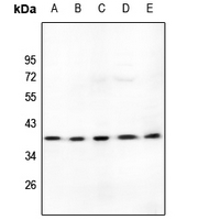 Western blot analysis of LDLRAD3 expression in HEK293T (A), A549 (B), PC12 (C), MEF (D), LO2 (E) whole cell lysates. (Predicted band size: 37 kD; Observed band size: 37 kD)
 -
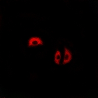 Immunofluorescent analysis of LDLRAD3 staining in A549 cells. Formalin-fixed cells were permeabilized with 0.1% Triton X-100 in TBS for 5-10 minutes and blocked with 3% BSA-PBS for 30 minutes at room temperature. Cells were probed with the primary antibody in 3% BSA-PBS and incubated overnight at 4 °C in a hidified chamber. Cells were washed with PBST and incubated with a AF594-conjugated secondary antibody (red) in PBS at room temperature in the dark.