
 Datasheet
 MSDS
MSDSDescription:Rabbit polyclonal antibody to ERK1Immunogen:KLH-conjugated synthetic peptide encompassing a sequence within the center region of human ERK1. The exact sequence is proprietary.Purification:The antibody was purified by immunogen affinity chromatography.Clonality:PolyclonalForm:Liquid in 0.42% Potassium phosphate, 0.87% Sodium chloride, pH 7.3, 30% glycerol, and 0.01% sodium azide.Dilution:WB (1/500 - 1/1000), IF/IC (1/100 - 1/500)Gene Symbol:MAPK3Alternative Names:ERK1; PRKM3; Mitogen-activated protein kinase 3; MAP kinase 3; MAPK 3; ERT2; Extracellular signal-regulated kinase 1; ERK-1; Insulin-stimulated MAP2 kinase; MAP kinase isoform p44; p44-MAPK; Microtubule-associated protein 2 kinase; p44-ERK1
Entrez Gene (Human):
5595;
Entrez Gene (Mouse):
26417;
Entrez Gene (Rat):
50689;
SwissProt (Human):
P27361;
SwissProt (Mouse):
Q63844;
SwissProt (Rat):
P21708;
Storage/Stability:Shipped at 4°C. Upon delivery aliquot and store at -20°C for one year. Avoid freeze/thaw cycles.
-
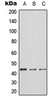 Western blot analysis of ERK1 expression in A431 (A), NIH3T3 (B), PC12 (C) whole cell lysates. (Predicted band size: 43 kD; Observed band size: 44 kD)
 -
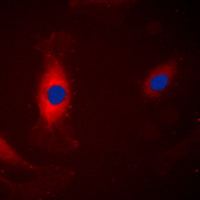 Immunofluorescent analysis of ERK1 staining in A431 cells. Formalin-fixed cells were permeabilized with 0.1% Triton X-100 in TBS for 5-10 minutes and blocked with 3% BSA-PBS for 30 minutes at room temperature. Cells were probed with the primary antibody in 3% BSA-PBS and incubated overnight at 4 °C in a humidified chamber. Cells were washed with PBST and incubated with a DyLight 594-conjugated secondary antibody (red) in PBS at room temperature in the dark. DAPI was used to stain the cell nuclei (blue).