
 Datasheet
 MSDS
MSDSDescription:Rabbit polyclonal antibody to MINA53Immunogen:KLH-conjugated synthetic peptide encompassing a sequence within the center region of human MINA53. The exact sequence is proprietary.Purification:The antibody was purified by immunogen affinity chromatography.Clonality:PolyclonalForm:Liquid in 0.42% Potassium phosphate, 0.87% Sodium chloride, pH 7.3, 30% glycerol, and 0.01% sodium azide.Dilution:WB (1/500 - 1/1000), IF/IC (1/50 - 1/200)Gene Symbol:MINAAlternative Names:MDIG; MINA53; NO52; Bifunctional lysine-specific demethylase and histidyl-hydroxylase MINA; 60S ribosomal protein L27a histidine hydroxylase; Histone lysine demethylase MINA; MYC-induced nuclear antigen; Mineral dust-induced gene protein; Nucleolar protein 52; Ribosomal oxygenase MINA; ROX
Entrez Gene (Human):
84864;
Entrez Gene (Mouse):
67014;
Entrez Gene (Rat):
266670;
SwissProt (Human):
Q8IUF8;
SwissProt (Mouse):
Q8CD15;
SwissProt (Rat):
Q8CFC1;
Storage/Stability:Shipped at 4°C. Upon delivery aliquot and store at -20°C for one year. Avoid freeze/thaw cycles.
-
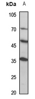 Western blot analysis of MINA53 expression in rat liver (A) whole cell lysates. (Predicted band size: 52 kD; Observed band size: 53 kD)
 -
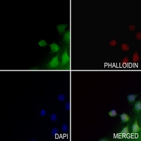 Immunofluorescent analysis of MINA53 staining in A549 cells. Formalin-fixed cells were permeabilized with 0.1% Triton X-100 in TBS for 5-10 minutes and blocked with 3% BSA-PBS for 30 minutes at room temperature. Cells were probed with the primary antibody in 3% BSA-PBS and incubated overnight at 4 °C in a hidified chamber. Cells were washed with PBST and incubated with a AF488-conjugated secondary antibody (green) in PBS at room temperature in the dark. Phalloidin - AF594 was used to stain Actin filaments (red). DAPI was used to stain the cell nuclei (blue).