
 Datasheet
 MSDS
MSDSDescription:Rabbit polyclonal antibody to ACSL1Immunogen:Recombinant fusion protein of human ACSL1. The exact sequence is proprietary.Purification:The antibody was purified by immunogen affinity chromatography.Clonality:PolyclonalForm:Liquid in 0.42% Potassium phosphate, 0.87% Sodium chloride, pH 7.3, 30% glycerol, and 0.01% sodium azide.Dilution:WB (1/500 - 1/2000), IF/IC (1/50 - 1/200)Gene Symbol:ACSL1Alternative Names:FACL1; FACL2; LACS; LACS1; LACS2; Long-chain-fatty-acid--CoA ligase 1; Acyl-CoA synthetase 1; ACS1; Long-chain acyl-CoA synthetase 1; LACS 1; Long-chain acyl-CoA synthetase 2; LACS 2; Long-chain fatty acid-CoA ligase 2; Palmitoyl-CoA ligase 1; Palmitoyl-CoA ligase 2
Entrez Gene (Human):
2180;
Entrez Gene (Mouse):
14081;
Entrez Gene (Rat):
25288;
SwissProt (Human):
P33121;
SwissProt (Mouse):
P41216;
SwissProt (Rat):
P18163;
Storage/Stability:Shipped at 4°C. Upon delivery aliquot and store at -20°C for one year. Avoid freeze/thaw cycles.
-
 Western blot analysis of ACSL1 expression in HepG2 (A), mouse liver (B), rat heart (C) whole cell lysates. (Predicted band size: 76; 77 kD; Observed band size: 78 kD)
 -
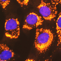 Immunofluorescent analysis of ACSL1 staining in NIH3T3 cells. Formalin-fixed cells were permeabilized with 0.1% Triton X-100 in TBS for 5-10 minutes and blocked with 3% BSA-PBS for 30 minutes at room temperature. Cells were probed with the primary antibody in 3% BSA-PBS and incubated overnight at 4 °C in a humidified chamber. Cells were washed with PBST and incubated with a AF594-conjugated secondary antibody (red) in PBS at room temperature in the dark. DAPI was used to stain the cell nuclei (blue).