
 Anti-LDHA Antibody
  Datasheet
 MSDS
MSDS
Description: Rabbit polyclonal antibody to LDHA Immunogen: Recombinant full length protein of human LDHA Purification: The antibody was purified by immunogen affinity chromatography. Clonality: Polyclonal Form: Liquid in 0.42% Potassium phosphate, 0.87% Sodium chloride, pH 7.3, 30% glycerol, and 0.01% sodium azide. Dilution: WB (1/500 - 1/2000), IF/IC (1/10 - 1/100) Gene Symbol: LDHA Alternative Names: L-lactate dehydrogenase A chain; LDH-A; Cell proliferation-inducing gene 19 protein; LDH muscle subunit; LDH-M; Renal carcinoma antigen NY-REN-59Entrez Gene (Human): 3939Entrez Gene (Mouse) : 16828Entrez Gene (Rat) : 24533SwissProt (Human): P00338SwissProt (Mouse) : P06151SwissProt (Rat) : P04642Storage/Stability : Shipped at 4°C. Upon delivery aliquot and store at -20°C for one year. Avoid freeze/thaw cycles.
-
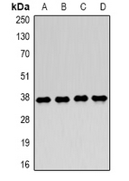 Western blot analysis of LDHA expression in MCF7 (A), Jurkat (B), NIH3T3 (C), mouse skeletal muscle (D) whole cell lysates.
 -
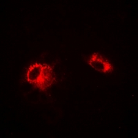 Immunofluorescent analysis of LDHA staining in A549 cells. Formalin-fixed cells were permeabilized with 0.1% Triton X-100 in TBS for 5-10 minutes and blocked with 3% BSA-PBS for 30 minutes at room temperature. Cells were probed with the primary antibody in 3% BSA-PBS and incubated overnight at 4 °C in a humidified chamber. Cells were washed with PBST and incubated with a DyLight 594-conjugated secondary antibody (red) in PBS at room temperature in the dark.
Differential Expression of Lonp1 Isoforms in Cancer Cells