
 Multiplex IHC Detection Kit (Triple)
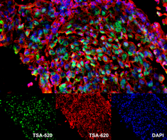
 Datasheet
 MSDS
MSDS
Description: Three colors multiplex IHC tyramide signal amplification (TSA) Directions for Use : 1. De-paraffinizing (de-waxing) and rehydrating
1.1 Heat the slides in tissue-drying oven for 45 minutes at 60°C. Place the slides in a rack, and perform the following washes:
Xylene I: 15 mins
Xylene II: 15 mins
100% Ethanol: 5 mins
95% Ethanol: 5 mins
85% Ethanol: 5 mins
70% Ethanol: 5 mins
1.2 The slides are placed in a lab draught cupboard to rinse off ethanol.
1.3 Finally, wash the slides in the pure water.
2. Antigen or epitope retrieval
2.1 Add the appropriate antigen retrieval buffer (EDTA pH 9.0 or sodium citrate pH 6.0) to the microwaveable vessel.
2.2 Place the slides in the microwaveable vessel. Then place the vessel inside the microwave (1200W).
2.3 Boil for 8 mins in microwave (1200 W) under medium heat, then stop heating for 8 mins, followed by low and medium heat for 7 mins. Other heat-induced epitope retrieval methods can also be used, e.g., heated at 120 °C 1-2 min, 100 °C 20mins or 95 °C in a water bath.
Be sure to monitor for evaporation and watch out for boiling over during the procedure. Do not let the slides dry out.
2.4 After cooling down in room temperature, place the slides in PBS (pH 7.4) to wash 3 X 5 mins on a decolorizing shaker.
Notes: To get best results, antigen retrieval buffer and protocol should be determined according to the tissue types and antigen types.
3. Blocking endogenous peroxidase
3.1 Add enough 3% hydrogen peroxide (H2O2) to cover the slides.
3.2 Incubate for 15 mins in the dark at room temperature.
3.3 Place the slides in PBS (pH 7.4) to wash 3 X 5 mins on a decolorizing shaker.
4. Blocking
4.1 Drain slides and then use an IHC pen to draw a circle around each sample on your slide (to hold antibody solution within the target area).
4.2 Add 3% BSA-PBST solution (or other blocking buffer) inside the circle to cover the tissues, incubate 30 mins at room temperature.
5. Primary antibody incubation
5.1 Remove blocking buffer and add primary antibody diluted by recommended antibody diluent overnight at 4°C or 37°C for 1-2h.
6. HRP Polymer Working Solution incubation
6.1 Place the slides in PBS (pH 7.4) and wash 3 X 5 mins on a decolorizing shaker.
6.2 Incubate slides with HRP Polymer Working Solution (50 μl for each slice) in the dark at room temperature for 60 mins.
6.3 Wash 3 X 5 mins with PBS buffer.
7. Tyramide labeling
7.1 Apply the TSA-520 Dye Working Solution to each sample (50 μl for each slice) and incubate for 10-15 mins at room temperature.
7.2 Wash 3 X 5 mins with PBS buffer.
8. Denoise
Repeat steps 2. At the end of the single stain, may add antifade mounting medium to view the slides or go on labeling another fluorescent dye.
9. Repeat
Repeat steps 3-7 for another fluorescent dye (TSA-620 Dye Working Solution) to each sample.
10. DAPI counterstaining
10.1 Place the slides in PBS (pH 7.4) and wash 3 X 5 mins on a decolorizing shaker.
10.2 Apply the DAPI solution (50 μl for each slice) to each sample and incubate in the dark at room temperature for 10 mins.
11. Mounting the slides
11.1 Place the slides in PBS (pH 7.4) and wash 3 X 5 mins on a decolorizing shaker.
11.2 Add antifade mounting medium (3-5 μl for each slice) to cover the section.
12. View the slides
View the sample using a fluorescence microscope with appropriate filters.
Note: To get the best results from multiplex staining, the experimental condition must be optimized.Application : Tyramide signal amplification (TSA), also called catalyzed reporter deposition (CARD), is a highly sensitive enzymatic method which can enable the detection of low-abundance targets in histochemical analysis. TSA utilizes the catalytic activity of HRP for the covalent deposition of labeled tyramide on and near target proteins or nucleic acid sequences in situ. In the presence of low hydrogen peroxide (H2O2), HRP is able to convert labeled tyramide substrates into highly-reactive, short-lived tyramide radicals that rapidly bind to tyrosine residues on and proximal to the enzyme site. These labels can be detected by standard chromogenic or fluorescent techniques. Multiple rounds of tyramide signal amplification can be performed for multicolor detection.Storage/Stability : Store at 4 °C in dark for 1 year, do not freeze.