
 DiR Membrane Staining Kit
  Datasheet
 MSDS
MSDS
Description: Highly deep red fluorescent membrane probe for labeling live and fixed cells Form: Liquid CAS Number : 100068-60-8Molecular Formula : C63H101IN2Molecular Weight : 1013.4Directions for Use : 1. For adherent cells staining
(1) Adherent cells were cultured on a sterile cover slide.
(2) Remove the cover glass from the medium, absorb excess liquid but keep the surface moist.
(3) Add 100 μL of Working Solution to one corner of the cover glass and gently shake to evenly cover all cells.
(4) Cells were incubated at 37 °C for 2-20 mins. The reaction time can be optimized to obtain uniform labeling effect.
(5) Discard Working Solution, wash the glass with PBS for 2 to 3 times.
2. For suspension cells staining
(1) Adding an appropriate volume of Working Solution to re-suspension cells, the density of the cells is 1× 10⁶ /mL.
(2) The cells were incubated at 37°C for 2-20 min. The reaction time can be optimized to obtain uniform labeling effect.
(3) After incubation, centrifuge at 1000-1500 rpm for 5 mins. Discard the supernatant and slowly add the growth medium again to resuspend the cells.
(4) Repeat step (3) more than twice.Components : The deep red fluorescent, lipophilic carbocyanine DiIC18(7) is widely used as a lipophilic tracer. It is weakly fluorescent in water, but highly fluorescent and quite photostable when incorporated into membranes. It has an extremely high extinction coefficient and short excited-state lifetimes in lipid environments. Once applied to cells, the dye diffuses laterally within the plasma membrane.Storage/Stability : Shipped at 4°C. Store at -20°C and protect from light for 12 months.
Platform : Ex/Em = 748/780 nm
-
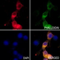 Immunofluorescent analysis of DiR staining in PC3 cells. Formalin-fixed cells were permeabilized with 0.1% Triton X-100 in TBS for 5-10 minutes and blocked with 3% BSA-PBS for 30 minutes at room temperature. Cells were probed with the primary antibody in 3% BSA-PBS and incubated overnight at 4 °C in a hidified chamber. Cells were washed with PBST and incubated with DiR (deep red) at room temperature in the dark. Phalloidin - AF488 was used to stain Actin filaments (green). DAPI was used to stain the cell nuclei (blue).