
 Hoechst 33342 Staining Kit
  Datasheet
 MSDS
MSDS
Description: Hoechst 33342 Staining Kit Application : Hoechst 33342 is a cell permeable fluorescent minor groove-binding probe for DNA, and specific stain for AT-rich regions of double-stranded DNA. This fluorescent dye has been used in sorting living cells, used in flow cytometry for the determination of DNA and used for visualization of chromatin distribution in living cells. Hoechst 33342 and DNA complex show light blue flourescent color with excitation light 355 nm and emission light 465 nm.Storage/Stability : Store at -20°C for one year.
-
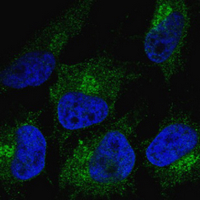 Immunofluorescent analysis staining in HeLa cells. Formalin-fixed cells were permeabilized with 0.1% Triton X-100 in TBS for 5-10 minutes and blocked with 3% BSA-PBS for 30 minutes at room temperature. Cells were probed with the primary antibody in 3% BSA-PBS and incubated overnight at 4 °C in a humidified chamber. Cells were washed with PBST and incubated with a FITC-conjugated secondary antibody (green) in PBS at room temperature in the dark. Hoechst 33342 was used to stain the cell nuclei (blue).